All Posts By

Mediterraneo Hospital

Mediterraneo Hospital 2000 - Mediterraneo Hospital: World First in Endoscopic Mastectomy Using the HandX® Robotic System

Mediterraneo Hospital: World First in Endoscopic Mastectomy Using the HandX® Robotic System

By MBC, PRESS RELEASES

PRESS RELEASE

Glyfada, 17th July 2025

 Mediterraneo Hospital: World First in Endoscopic Mastectomy Using the HandX® Robotic System

Mediterraneo Hospital announces the world’s first successful endoscopic mastectomy using the innovative HandX® robotic system, marking a new era in minimally invasive surgical treatment of breast cancer.

The pioneering procedure was performed by the Breast Clinic surgical team, led by Clinical Director Dr. Vasileios Kalles, in collaboration with Deputy Director Dr. Ioannis Papapanagiotou, Gynecologist – Breast Surgeon, and Dr. Apostolos Mitrousias, Breast Surgeon.

With an incision of only 3 centimeters, the technique allows for the simultaneous removal and reconstruction of the breast with exceptional precision, reducing surgical trauma and offering faster recovery and improved aesthetic results for the patient, compared to traditional mastectomy methods.

The HandX® system provides enhanced dexterity and control, enabling the safe and effective application of endoscopic mastectomy. This internationally pioneering procedure, performed for the first time at Mediterraneo Hospital, demonstrates the hospital’s commitment to innovation and to providing high-quality, personalized care.

Dr. Vasileios Kalles commented:
“We are proud to be at the forefront of surgical innovation. By combining endoscopic access with robotic precision, we are redefining the possibilities in breast cancer surgery. This new technique brings significant benefits to patients, and we are particularly pleased to offer such advanced surgical methods in Greece and at Mediterraneo Hospital’s Breast Clinic.”

This pioneering procedure reflects the Breast Clinic’s dedication to providing high-level services through an interdisciplinary approach and the integration of the most advanced techniques for breast cancer treatment and prevention.
At the same time, it serves as another example of Mediterraneo Hospital’s firm orientation towards technological innovation, driven by the commitment to place the patient at the center of care.

mediterraneo hospital ski snowboard vouno trafmatismoi - Stay safe on the slopes!

Stay safe on the slopes!

By ATHLETIC TRAUMA, IN THE NEWS, NEUROSURGERY

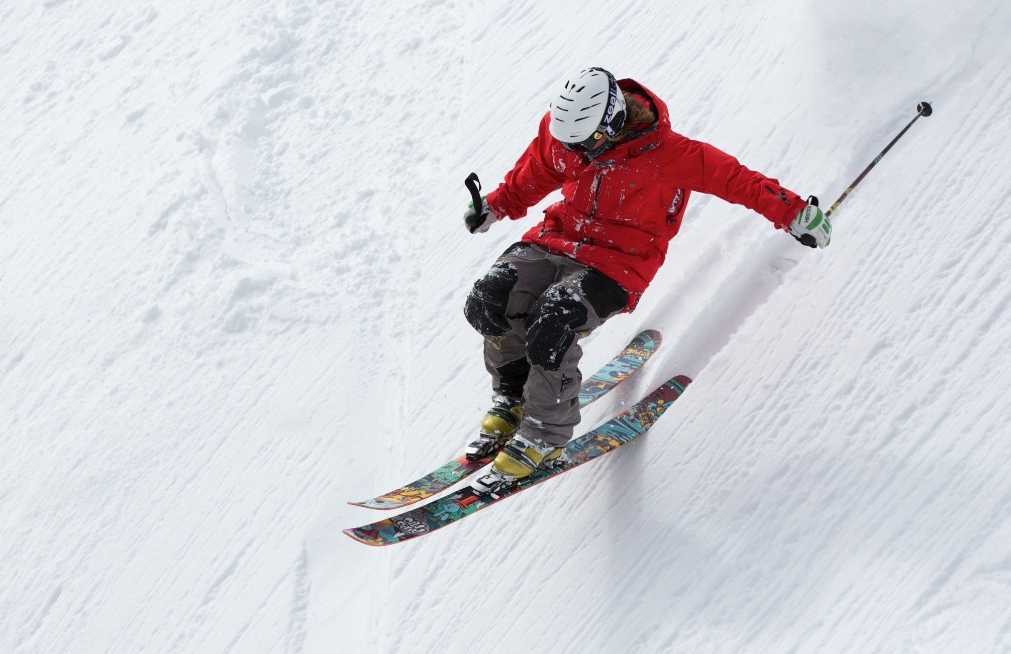
Skiing and snowboarding are exciting winter sports, but they entail risk, especially for the head and spine. Falls and collisions are the main causes of injuries. It is estimated that 1.5 to 6 injuries per 1,000 skiing days. Injuries to the lower and upper extremities are the most common, but head and spine injuries have the most serious consequences. The case of M. Schumacher, who suffered a severe head injury while skiing in December 2013, is very characteristic.

stavropoulos mediterraneo hospital - Stay safe on the slopes!

Dr. Stavros Stavropoulos, Neurosurgeon
Registar, Mediterraneo Hospital Neurosurgery Dpt.

Head injuries can initially be underestimated. Moreoften than not, a brain injury develops later in the day or on the next day. The symptoms of a head injury should never be ignored. Even in the case of minimal symptoms and a mild concussion, the discomfort can lead to a second, potentially tragic, accident.

Therefore, if you happen to suffer a head injury, it is best to stay safe and avoid further exposure to unnecessary danger. Spinal cord injuries, on the other hand, may require immediate immobilization and transport to a medical facility following all of the appropriate transport protocols which may include immobilization on a stretcher and a neck collar right from the scene of the accident. Neurological complications that occur later on after an initial injury and are caused by improper handling of the injured person can even lead to paralysis. If you suspect you have a serious spinal cord injury – experiencing excessive pain in the spine or weak/numb legs or arms- remain still or minimize movement and seek medical care immediately.

Completely eliminating the risk of injury is only possible if you stay home. A more realistic approach would be to reduce the risk of injury, and this can be easily achieved by following a few simple tips:

  • Wear Protective Gear: Helmets significantly reduce the risk of head injury, without increasing the risk of neck injury.
  • Warm up: Stretching before starting a sports activity improves flexibility and reduces muscle strain.
  • Check your Equipment: Make sure your skis, bindings, and helmets are in good condition.
  • Know your Limits: Stay on a slope that matches your skill level to avoid exhaustion.
  • Stay Hydrated: Dehydration can affect concentration and increase fatigue.
  • Stay in Shape: Regular neuromuscular training with an emphasis on flexibility and balance helps prevent injuries.
  • Take Lessons: Certified instructors do teach proper technique, reducing the possibility of a fall.
  • Remember: Alcohol and ice only mix well in a glass, never on the slopes. Never on the slopes
cest tsoukas - Echocardiography Education with Simulation Training

Echocardiography Education with Simulation Training

By CARDIOLOGY, IN THE NEWS, SEMINARS

a.tsoukas 180x300 - Echocardiography Education with Simulation Training

Tsoukas Athanasios MD, PhD, FESC
Dir. Echo Lab Mediterraneo Hospital

Educational curricula in Echocardiography traditionally include theoretical knowledge and practical hands-on experience, initially obtained by performing supervised studies on real patients in a clinical setting. Competence in echocardiography requires a deep understanding of cardiac anatomy, physiology, pathophysiology and haemodynamics, which must then be correlated to the corresponding ultrasound images as well as technical aspects including ultrasonic physics. The learning curve for a trainee to become skilled and self-sufficient, takes a long time.

Moreover, experience requires familiarisation with various clinical scenarios and cardiac conditions, which is lengthening the duration of the procedures; it is accompanied by reduced productivity, potential patient safety concerns and considerable stress for the patient and the fellow in training. Nowadays there is a strong recommendation for using simulators in clinical practice. Echocardiography skills should be performed and learned in a simulation environment prior to undertaking them on real patients. Simulators is a modern way to learn normal and abnormal cardiac anatomy and function in a safe digital environment, promoting experience with safety of the patients.

Education using simulators customary in certain fields including aviation, space exploration, Formula 1 and Moto GP drivers for training purposes in complex settings and procedures. This method has been postulated as having the potential to assist trainees develop technical skills to expert level before undertaking them in reality, providing a safe environment in which they are allowed to make mistakes and learn from them, review performance and have the ability to stop the procedure and restart any parts of the training necessary.

Advantages include elimination of trainee stress, elongation of the time procedure as needed particularly in techniques such as transesophageal echocardiography that require practical skills, acquisition of optimum quality images, practice in a variety of clinical cases and ongoing education. Trainees can perform a complete echo study with measurements and calculations, inserting all data and issuing a final report at the end of the examination. Logistics of the system permit for every trainee doctor to complete a personal logbook with all his studies on the simulator, for further evaluation and accreditation in the future.

Simulation training is not new to medicine. Anatomical models were created for practice in cardiopulmonary resuscitation. The first cardiovascular simulators were based on computer technology and provided cardiac anatomy with palpable pulse and areas for auscultation. These allowed medical students to experience some of the findings from clinical examination for first time in a mannequin.  Modern technology allows the creation of complex anatomical, physiological and pathophysiological systems programmed to respond to the user. Specifically in the field of Cardiology, a number of simulators for echocardiography, coronary angioplasty, pacing, transcatheter aortic valve replacement and peripheral vascular interventions are available.

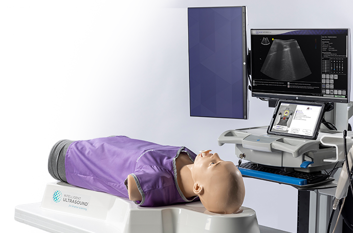
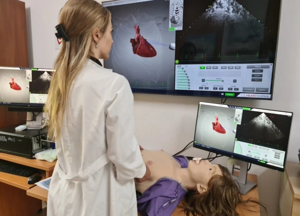
Echocardiography simulators are used both in Transthoracic and Transesophageal Imaging. Simulation-based learning offers to the trainee a high quality examination, in a low stress and low pressure environment with a functioning realistic probe and a realistic mannequin similar to living patients. The simulator enables a real time 3D reconstruction of the cardiac anatomy in a variety of clinical scenarios. In every case there is a simultaneous anatomically accurate and interactive 3D heart, so trainees can relate the anatomic to the ultrasound imaging. This 3D heart model can be rotated and sliced in any direction and the intracardiac structures can be highlighted (5).

Realistic transthoracic (TTE) scanning can be obtained and users can learn how to perform the standard echocardiographic exams in sequential images similarly to a real patient exam. As in ultrasound machines, simulators have modules for M-mode, biplane, colour, pulsed wave and continuous wave doppler in all regions. A software package for measurements and calculations is also available online.
2000 63dfda226a892 1024x738 - Echocardiography Education with Simulation Training
The system includes the gold standard imaging views for each of transesophageal (TEE) planes, side by side with 3D cardiac anatomy images, using the realistic TEE probe with controls for ante and retroflexion, lateral flexion and omniplane rotation.

Training is obtained in a wide range of clinical cases, including normal heart and lungs, various disease states and key cardiac devices. Coronary artery disease, valvular diseases, cardiomyopathies, pericardial diseases, cardiac tamponade, congenital heart disease, acute aortic syndromes, prosthetic valves, cardiac devices, pulmonary embolism, pneumothorax, as well as o lot of clinical cases and scenarios are included in the educational program of the simulation system (6). Testing platform allows tutors to create standardized or customized tests for students and is an effective assessment tool that can easily adapted to any training need.

Simulation equipment and modules alone cannot offer a complete education. Even tutors will require specialized training in equipment use and techniques, which requires skills not necessary for other forms of teaching. They need support from experienced educators to run successful simulation programs. The technology of simulation is rapidly progressing, offering last generation and high fidelity devices, more cardiac diseases and clinical scenarios, and advanced modules including current minimally invasive procedures, such as TAVI, mitral clip, left atrium appendage occlusion with device, lung scan in Covid patients.

Additionally there is a growing simulation faculty, with national and international societies, conferences and peer reviewed publications. American Society of Echocardiography and European Society of Cardiovascular Imaging schedule training recommendations to standardize the use of these new learning and training methods, separately from the existed guidelines for Transesophageal and Transthorasic Echocardiography.

Simulation in Cardiology and Echocardiography is expanding rapidly due to advances in digital technology. A new way of training is now available in a safe environment, without any risk for the patient, extended training hours and ability for more progressive skill acquisition and maintenance. In the near future, possibly before any healthcare professional performs a skill on a patient, should have the opportunity to perform the same skill in a simulation device. This is especially important as exposure to patients by junior doctors and training hours are reduced. Simulation assures that cardiologists in training have reached the minimum standards of competence before performing procedures on patients as well as experienced operators further specialize in new technologies.

In conclusion, simulators in Echocardiography offer a full education program in Transthoracic (TTE) and Transesophageal Echocardiography (TEE) for trainees in Cardiology, Cardiologists, Anesthesiologists, and all doctors who are interest in the field of Echocardiography. The software provides side-by-side best quality 3D anatomical images, echo views in virtual reality and a wide range of cardiac diseases. Simulator allows realistic probe positioning for both TTE and TEE echo. TEE probe offers all the authentic controls of a real probe and accurate enables real time anatomical and ultrasound views. Colour, PW and CW Doppler modalities, 2D and Doppler measurements are also available with the tools for a quantitative assessment. Technology in simulation is rapidly progresses and modules for 3D studies are now under comprehensive evaluation, and will be suitable for use in the daily clinical practice at the end of this year.

ski dr.patsopoulos - Ski Safety

Ski Safety

By ATHLETIC TRAUMA, IN THE NEWS, ORTHOPAEDICS

hercules patsopoulos mediterraneo hospital - Ski SafetyM.D. Heracles I. Patsopoulos, Orthopaedic Surgeon, Head of the Athletic Trauma Clinic, Mediterraneo Hospital


What we need to be careful for during activities in the mountain and the ski centre.

According to recent data by the American Academy of Orthopaedic Surgeons, every year 15% of those taking on skiing are injured and transferred to health clinics, while statistically it has been observed that 70% of these injuries happen at the end of a day. Tiredness often seems to be the cause behind this, since people go the extra mile to finish up their work out. During the last years, there has been a great increase in snowboard related injuries. If an injury occurs, involving a fall or even without one, the winter athlete needs to stop their activity, even if they don’t feel acute pain, since the body is still warm. They need to rest for at least half a day and if the pain persists they need to seek ot medical advice.

The most usual injuries are meniscus tears, tears of the ligaments of the anterior cruciate ligament and the medial collateral ligament, injuries of the spinal cord, pelvis fractures, dislocations and fractures of the shoulder, the elbow and the fingers and toes, as well as multiple strains and tears of muscles and tendons, which constitute slightly less serious damage. Burns due to friction are also often. The most important damage are craniocerebral injuries which also have the worst prognosis. The most usual snowboard injuries are injuries at the wrist, while the most usual skiing injuries have to do with the lower limbs and mainly the anterior cruciate ligament.

Head injuries are approximately 28% of skiing injuries and 33.5% of snowboarding injuries. In total, 22% of head trauma is serious enough to cause clinical symptoms of concussion, while 88% of all deaths occurring following skiing and snowboarding accidents are due to craniocerebral injuries. Fractures of the hip are more serious, while common among people over the age of 60 due to osteoporosis, and they call for hospital admission and surgical operation.

The low temperature causes muscles and blood vessels to contract, lowers stamina and increases the possibilities of painful muscle contraction which increases the danger of injuries. An important precautionary measure is warm up before engaging in any athletic activity in the snow, which needs to be followed by a simple stretching routine so that the muscles are not “cold” when you start going downhill on any means. If such a precaution is not taken, the chance for ligament or muscle strains is great. The speed at which one slides down the snow or the intensity with which one stops their course downhill when skiing demand great muscle strength and ligament stability.

Appropriate equipment in every sport constitutes the cornerstone in preventing injuries, foremost among which is the use of a helmet, as shown in the above. An overview of the literature regarding the efficiency of the use of a helmet in mitigating head injuries in snow sport athletes proved the unequivocal priority of the use of a helmet in mitigating risk as well as the severity of cranial injuries in said athletes. The use of a helmet should be MANDATORY for children. Ideally, the helmet needs to be aligned with the European (EN 1077) or American (SnellRS98 or ASTMF2040) standards.

Modern ski boots are designed to provide exceptional control and support, therefore limiting the range of movement of the ankle joint. Ski straps are now adjustable depending on the weight, the height, the age and the speed and they are calibrated at the beginning of each season. A tight ski strap (which fails to be released during a usual fall) leads to a tear of the anterior cruciate ligament three times more often in comparison to a more lax ski strap. Falling at a low speed and backwards results in a higher chance of ski straps not being released.

Snowboard straps, contrary to those of recreational skiing, are not releasable, while there are two main types of snowboard boots: the soft ones and the hard ones. Each boot type exerts different forces upon the foot and the lower limp and many studies have shown that, when snowboarding, 50% of injuries at the lower limps are associated with the type of boot used. Hard boots expose snowboarders to the risk of a fractured shin and fibula at the top of the boot (know as boot top fracture) and double the risk of a knee injury as compared to soft boots. However, soft boots expose snowboarders to double the risk of ankle injury as compared to hard boots.

The rules to follow unfailingly when moving outside the pistes of the ski centre are:

  • Your outing should take place around noon and not in the morning or at sun-down to avoid frost (if possible).
  • You should always wear double socks, a beanie, gloves, dry and thermal clothes (if available).
  • You should always wear appropriate boots and you should never wear sneakers or shoes with leather soles, as these are highly slippery on ice.
  • You should walk with slightly open legs to expand our supporting base.
  • You shouldn’t walk fast, but at half your average step.
  • You should never walk with your hands in your pockets, since, in case you slip, you need to be able immediately and efficiently correct your slip and restore your centre of mass over your supporting base.
  • You should never run on snow or ice.
  • You shouldn’t walk on ice or near cars passing by since there is a danger of injury.
  • You should always try to step on fresh snow.
  • You should avoid a steep route uphill or downhill as well as stairs, whenever possible. When one falls from an upright position forward, their first reflex is to put their hands first in order to protect themselves. Therefore, they fall hands and knees first, possibly causing injuries ranging from a simple lesion to fractures. A broken bone is extremely painful, it is obviously deformed and on occasion it can give the patient a sense of odd mobility of the limb. If you cannot avoid falling, you should try and fall on your side, rolling, in order to mitigate and better control the momentum you have while falling. This particular type of fall is more common with motorcycle riders and you need but to watch a few such videos of falls.For all those who love winter sports, preparation should start at least one month beforehand, with exercises to strengthen your abdominal and back muscles as well as your quadriceps being essential for your protection against injuries on the mountain. Despite our advice, accidents can happen at any moment, therefore you should remember that the only one not having accidents on the mountain and the snow is the one who never left the house.

Source: Snowflake Magazine

CMR031 Certificate V2 1 - European CMR Laboratory Accreditation for the Cardiac MRI Unit of Mediterraneo Hospital

European CMR Laboratory Accreditation for the Cardiac MRI Unit of Mediterraneo Hospital

By CARDIOLOGY, IN THE NEWS, MRI

CMR031 Certificate V2 1 1024x724 - European CMR Laboratory Accreditation for the Cardiac MRI Unit of Mediterraneo Hospital

We are pleased to announce that the Cardiac MRI Unit of Mediterraneo Hospital has been recently accredited by the European Association of Cardiovascular Imaging (EACVI), member of the European Society of Cardiology (ESC).

This is the first and only accredited Cardiac MRI Unit in Greece up to date, and one of the 21 accredited Cardiac MRI Units in Europe. This distinction comes as a recognition of the high-quality services that are provided at the Cardiac MRI Unit at Mediterraneo Hospital.

The MRI Cardiac Unit of Mediterraneo Hospital is one of the largest in Greece, with a total of approximately 700 studies annually, which cover the whole spectrum of cardiovascular diseases in a variable age spectrum of cardiological patients.

Our unit uses ultramodern technical equipment which consists of two state-of-the-art technology MRI scanners (Philips Ingenia 1.5T and 3T).

Our team consists of Radiologists with a vast experience in Cardiovascular Imaging, highly-trained dedicated Cardiac MRI radiographers, as well as a specialized nurse and paramedical staff, specifically trained in the preparation and handing of our patients.

Our chief Cardiologist is Dr. Maria Boutsikou MD,MSc,PhD, FESC, FSCMR, specialized in Cardiac MRI. Dr Boutsikou has been trained in Cardiac MRI Imaging of Adults and Children at the Royal Brompton Hospital in London and is the only Cardiologist in Greece up to date to hold all three following higher Certifications:

  • Certification Level 3 in MRI Cardiac Imaging, by the European Association of Cardiovascular Imaging- EACVI) member of the European Society of Cardiology- ESC).
  • Certification Level 3 in MRI Cardiac for children and patients suffering from congenital heart disease, by the European Association of Cardiovascular Imaging- EACVI) member of the European Society of Cardiology- ESC).
  • Certification level 3 in MRI Cardiac imaging by the Society for Cardiovascular Magnetic Resonance- SCMR).

This exceptional distinction certifies the ability of the Unit to incorporate the international Guidelines in the process and diagnosis of the Cardiac MRI studies and therefore provide high quality services to our patients.

Moreover, it solidifies the ability of the Unit to support and conduct an extensive Educational Programme  on Cardiovascular imaging and expand our present research collaborations with acclaimed reference Centers across Europe and USA.

dimitrios giotikas orthopaedic surgeon - Limb Reconstruction and Lengthening

Limb Reconstruction and Lengthening

By IN THE NEWS, ORTHOPAEDICS
dimitrios giotikas orthopaedic surgeon - Limb Reconstruction and Lengthening

Dr. Dimitrios M. Giotikas, Orthopaedic Surgeon, Mediterraneo Hospital

Limb reconstruction of the upper and lower limbs (Limb reconstruction) is the field of orthopaedics that specializes in treatments related to bridging bone defects, correcting bone deformities and lengthening of the limbs; or any combination of the above.

These methods are applied to patients with:

  • Bone infections after a fracture or surgery; Chronic osteomyelitis.
  • Delayed Union, non-union or pseudarthrosis of fractures (inability to heal/unite).
  • Severe open fractures with loss of bone, skin and muscles.
  • Deformities of the limb (bending, shortening, rotational or any combination thereof) after a fracture or complication of previous surgery.
  • Joint stiffness – Knee and ankle joint fixed stiffness and ankylosis.
  • Congenital abnormalities, Leg Length discrepancy, limb hypoplasia, dwarfism.
  • Short stature, height dysphoria; cosmetic limb lengthening

These problems usually come as a result of the severity of the initial injury and/or as complications that occurred during its subsequent treatment. The social, professional and personal life of the patient is significantly affected for prolonged period (Life-changing events).

Dealing with these problems is difficult and time consuming. Often, the input of many specialties is required (Orthopaedics and Plastics Surgeons, Infectious Diseases Specialist, Physiotherapist, Prosthetics Orthotist, Psychologist).

giotikas dim - Limb Reconstruction and Lengthening
Our medical team consists of specialized doctors with substantial clinical experience from large international tertiary trauma centres.

In our department we use cutting-edge technologies such as:

  • Magnetically expandable intramedullary nails (Precise-2®, Nuvasive) for femoral and tibial lengthening with or without deformity correction.
  • Mechanically expandable intramedullary nails (Guichet X-OS®) that allow walking with a full weight bearing from the first postoperative day.
  • Circular external fixators for multi-plane correction of complex limb deformities using special software (Taylor Spatial Frame®, Smith & Nephew).
  • Special intramedullary reamer (Reamer Irrigator Aspirator RIA® by Johnson & Johnson) for obtaining bone graft with minimal invasive technique and also for intramedullary debridement and lavage in cases of extended intramedullary osteomyelitis.
  • Absorbable antibiotic-carrying pellets (Cerament® Bonesupport and Stimulan® Biocomposites) for the local adjuvant treatment of bone infections.

Our team gets referrals of patients from around the world, whose complex orthopedic problems require specialized surgical expertise  and the most advanced medical technology.

alexandra nikita mediterraneo hospital - Breast MRI

Breast MRI

By IN THE NEWS, MBC, MRI

Dr. Alexandra Nikita, Consultant Breast Radiologist, Mediterraneo Hospital

Breast MRI is a high-resolution imaging test for the examination of breast structures and is performed in addition to a mammography and breast ultrasound. It is a short, completely painless and harmless diagnostic test, during which the examinee does not receive any radiation.

In which cases is breast MRI indicated?

Breast MRI does not replace a mammography or breast ultrasound but is a complementary tool used in the following cases:

  • Screening in women at high risk of developing breast cancer. This includes women with a family history of breast or ovarian cancer (mother or sister who developed breast cancer before the age of 50, or more than one close relatives, including those on the examinee’s father side, with a history of breast or ovarian cancer),
  • Women with BRCA1 (Breast Cancer Type 1 susceptibility gene) and BRCA2 (Breast Cancer Type 2 susceptibility gene) mutations and women with a history of chest radiation therapy at the age of 10-30 years old. In any case, your radiologist or breast surgeon will review your family history and determine if a breast magnetic resonance imaging is appropriate for you.
  • To further investigate equivocal mammography findings. In rare cases, if a lesion detected on a mammogram cannot be adequately evaluated by MRI-guided localization, tomography, or ultrasound, magnetic resonance imaging is used to determine whether or not a biopsy is necessary,
  • To accurately determine the extent of the lesion in specific types of diagnosed cancer and exclude the presence of other foci in the same or the other breast, especially in dense breasts and in cases of young women with breast cancer. Breast MRI also investigates the possible presence of abnormal axillary lymph nodes,
  • In cases of a previous breast cancer surgery to investigate possible recurrence or development of new foci in the same or the other breast (method of choice),
  • To evaluate the mastectomy area after cancer treatment. Surgery scars and recurrent cancer may present some similarities on a mammogram and breast ultrasound. If a change in the mastectomy scar is detected, either via mammography or physical examination, magnetic resonance imaging can show if it is scar tissue or cancer recurrence,
  • To assess the response of the tumor to chemotherapy in cases where preoperative chemotherapy is necessary (neoadjuvant chemotherapy),
  • For the evaluation of breast implants and the investigation of their rupture.

mri mediterraneo hospital - Breast MRI

How is a breast MRI performed?

The examination is performed on an outpatient basis and lasts about 30′. The examinee is  placed in a prone position on the specially designed MRI scanner.

The comfort of the examinee during the whole process is important, because in that way she can remain calm and immobile for as long as necessary in order to complete the examination successfully.
The use of an intravenous contrast media (IV contrast) is necessary to investigate possible malignancy, because without it the examination presents insufficient results. If, however, the breast MRI is performed only to evaluate the breast implants, IV contrast is not required except in cases where inflammation in the area of the implant is investigated.

What are the disadvantages of a breast MRI?

The method has two main disadvantages: The first is the high rate of “false-positive” results, i.e. when the MRI reveals pathological findings that prove to be benign formations or areas of active glandular tissue. This is proven by a negative biopsy. The second disadvantage is the inability of the method to detect breast microcalcifications, suspicious for onset malignancy.

DIMOU 800Χ600 - 6 ways to treat chronic low back pain without surgery

6 ways to treat chronic low back pain without surgery

By CENTER FOR PAIN MANAGEMENT, IN THE NEWS

Chronic back pain is defined as pain that persists for 12 weeks or more, even after an initial injury or acute back pain has been treated. About 20 percent of people with acute back pain develop chronic back pain with persistent symptoms within a year. Even if the pain persists, it does not always mean a medically underlying severe cause or a cause that can be easily identified and treated. In some cases, treatment successfully relieves chronic back pain. Still, in other cases, the pain persists despite medical and surgical treatment.

Common causes of chronic back pain

Chronic back pain is usually associated with advanced age. However, it may also be due to a previous injury. The most common causes of chronic back pain are:

  • vertebral arthritis, i.e., the gradual destruction of cartilage between the joints of the spine.
  • spinal stenosis, i.e., the narrowing of a part of the spinal canal that can cause pain due to pressure on the nerves in the area.
  • damage to the intervertebral discs (disc herniation, intervertebral disc degeneration).
  • myoperitoneal pain syndrome.

In some cases, it is difficult to identify the cause of chronic back pain. Suppose the source of the pain is not known or cannot be treated. In that case, the best option is for the patient to work with the doctor to reduce the pain’s exacerbations and treat it with non-surgical treatments.

Non-surgical treatments for chronic back pain

  1. Injectable treatments

They are used when the source of the pain is known and sometimes help rule out specific causes of back pain if the treatment does not work. Minimally invasive treatments include epidural steroid infusion, intrathecal steroid infusion, and transdermal neurolysis using radiofrequency ablation.

Injectable steroids are artificial synthetic drugs similar to cortisol, a natural hormone produced in the adrenal glands and brain. Steroids help reduce pain and inflammation and are used to treat various inflammatory diseases and painful conditions, including intervertebral disc herniation.

Epidural steroid infusion is a low-dose injection of anti-inflammatory drugs and analgesics into the lower back (waist) to relieve pain in the legs or abdomen. The drug is injected into an area called the epidural space. By reducing inflammation, epidural steroid injections can significantly help reduce pain. Epidural steroid infusion can be a treatment and a way of differentially diagnosing the source of pain in the lumbar structures when in doubt.

On the other hand, intrathecal injection of steroids is the direct injection of the same substances into the nerve foramen, i.e., at the point of exit of the nerve root from the spine.

Also, transdermal neurolysis using radiofrequency is an effective method of treatment. Many modern studies support reducing or even disappearing back pain after radiofrequency ablation (RFA) of the tiny sensory nerves located near the vertebral joints. It is a minimally invasive method aimed at destroying nerve cells by applying an electric field on a tissue. In this way, denervation is achieved (by cauterization or cold field), with consequent pain relief. Transdermal neurolysis using radio frequencies has evolved dramatically in recent years.

Injectable treatment can lead to remission or even elimination of pain for some time. Still, it is not a permanent solution and is used in combination with other therapies.

  1. Pharmacological treatments

Analgesics, anti-inflammatory drugs, muscle relaxants and other medications can be used to control chronic back pain. However, most are accompanied by side effects and are not intended for long-term use. Opioid analgesics are generally not recommended as monotherapy or as the first treatment for a patient before trying other medicines. It is also not recommended to be used as a long-term treatment. Many of them are addictive and do not address the underlying cause of back pain. They should only be prescribed after a thorough examination by a specialist and if other medicines have not been shown to be effective for the patient. In recent years there have been many clinical studies on the beneficial effects of natural anti-inflammatory drugs. It is essential that health professionals, especially those involved in the treatment and management of chronic pain, be fully informed on the various dietary supplements available to form a successful treatment plan with or without conventional medicine.

  1. Physiotherapy

Exercise is the foundation for treating chronic back pain. It is one of the first treatments that the patient should try under the guidance of a doctor and physiotherapist with experience in the spine. However, the same exercises are not suitable for all patients. The exercises should be individualized and adapted according to the patient’s symptoms and condition. It also seems important to maintain an exercise routine at home for the success of the treatment.

Physiotherapy for chronic back pain may include:

  • training to maintain proper posture
  • testing of pain tolerance limits
  • Stretching exercises and flexibility exercises
  • Aerobic Exercise
  • trunk strengthening
  1. Alternative therapies

Acupuncture, massage, biofeedback therapy, laser therapy, electrical nerve stimulation and other non-surgical treatments for the spine can also help treat chronic back pain.

  1. Nutrition

Some diets favor inflammatory processes in the body, especially those high in fat, processed carbohydrates and processed foods. Consult your doctor to see if your diet contributes to chronic back pain and how you can change it. Maintaining a healthy weight could also help reduce back pain by reducing the strain on the spine.

  1. Lifestyle change

The patient with chronic back pain must accept certain limitations in their daily life and adapt. He must learn to “listen” to their body and adjust the rhythm accordingly to daily activities. It is also good to observe which activities may aggravate the pain and to avoid them as much as possible. This can help reduce pain but also prevent the progression of the underlying pain-related condition. Another necessary lifestyle change is smoking cessation. Nicotine has been shown to aggravate pain and delay its healing.

When is surgery for low back pain indicated?

The following are warning signs that surgery is needed if they are found to be related to the condition of the spine:

  • new or worsening bowel or bladder disorder
  • weakness of the limbs
  • gait and balance disorders
  • signs of increased reflexes

Surgery may also be a good option in treating chronic back pain if the cause of pain is known and confirmed by imaging techniques or if other treatments have not helped.

reflux 1 - Gastro-Esophageal Reflux Disease and its treatment

Gastro-Esophageal Reflux Disease and its treatment

By GASTROINTESTINAL, IN THE NEWS

michail kourkoulos 300x300 - Gastro-Esophageal Reflux Disease and its treatmentDr Michail S Kourkoulos MSc FRCs, Laparoscopic GastroIntestinal and Bariatric Surgeon

Director of Upper GastroIntestinal and Antireflux Surgery, Mediterraneo Hospital


Gastro-Esophageal Reflux Disease (GERD) or Acid Reflux, is a chronic condition affecting 10-20% of the general population. It’s usually treated with diet and lifestyle modification, although frequently requiring further treatment with medication, and occasionally either endoscopic or surgical treatment methods. Below are the usual questions asked by GERD sufferers.

Which symptoms are suggestive of Gastro-esophageal reflux disease?

GERD presents with a variety of symptoms, caused by the pathological entry of gastric contents into the chest, to the point of mimicking coronary disease. It can present as regurgitation of food up to the mouth, or frequent vomiting. There can be respiratory problems or worsening asthma due to reflux of acid into the lungs. It can also cause halitosis and tooth decay due to chronic contact with acidic gastric contents. Most of these symptoms are worse at night due to sleeping horizontally, and after large meals.

What is the cause of GERD?

The most common cause is the presence of a hiatal hernia, which means that part of the stomach, along with the lower esophageal valve mechanism which under normal circumstances prevents reflux, is lying in the wrong anatomical part of the body, within the chest instead of the abdomen. Other causes include obesity, pregnancy which causes hormonal relaxation of the valve mechanism, medication such as cortisone and substances that lead to relaxation of the lower esophageal sphincter, as well as rare syndromes which cause either hyperacidity of the stomach, or esophageal dysfunction. GERD is usually worsened by increased body weight, eating habits and lifestyle. Symptoms can therefore be improved by weight reduction and lifestyle modification according to specialist guidance.

Is Gastro-Esophageal Reflux Disease dangerous?

Long term exposure of the esophagus to gastric acid can cause a wide range of problems. Initially there is chronic inflammation of the surface of the esophagus called esophagitis. If this is not dealt with, chronic intense inflammation can lead to stricture of the esophagus due to scarring, presenting as difficulty in swallowing solid foods. The most worrying complication of GERD is the development of Barrett’s Esophagus, a metaplastic transformation of the cells of the lining of the lower esophagus to another type which is also benign, but has a chance of malignant transformation and development of Lower Esophageal Adenocarcinoma if the symptoms are ignored.

What are the necessary dietary and lifestyle changes which help improve symptoms and reduce the possibility of further problems?

Weight loss is the most important change which leads to improvement of GERD symptoms. Avoidance of triggering foods is also very important. Coffee and chocolate contain substances which lead to relaxation of the lower esophageal sphincter, and avoiding them improves symptoms. The same applies for smoking. Fizzy drinks and beer expand the stomach due to release of CO2 gas, and increase the intragastric pressure causing the gastro-esophageal valve mechanism to open leading to worsening symptoms and therefore have to be avoided. Red wine also leads to reflux due to the flavonoids contained. It is also advised to avoid large meals, which once again lead to reflux via gastric distention. Spicy meals and certain foods such as tomato cheese and occasionally even apples worsen symptoms. It is advised to leave at least 3 hours between dinner and sleep, as a full stomach in a horizontal position will certainly cause reflux. Many patients use 2-3 pillows as a dependent position utilizes gravity to reduce reflux, although these tend to slip away during the night. My personal preference is elevating the top of the bed with either supports under the mattress or under the legs of the bed. Alternatively, there are purpose made wedge pillows that can be used.

Is there some form of treatment for improving symptoms until lifestyle changes take effect?

First line treatment for GERD symptoms consists of medications which reduce acidity of gastric contents, neutralize gastric acid, or protect the surface of the esophagus. The type, dosage and duration of treatment as well as any further investigation prior to its commencement, depend on the type, severity and duration of symptoms. Mild typical symptoms which improve with lifestyle changes alone do not require any further investigation, although this decision is best made by a specialist. The most common first line treatment is a 4–6-week trial of Proton Pump Inhibitors such as Omeprazole, Lansoprazole, Esomeprazole etc., alone or in combination with Gaviscon. In case of side effects, alternative medication can be used according to doctor’s guidance. Medical management reduces the acidity of refluxate and acid exposure of the esophagus, but does not treat the cause of reflux, which is dysfunction of the valve mechanism of the lower esophagus.

What happens if symptoms recur as soon as treatment stops?

Indeed, in many cases GERD symptoms reappear as soon as the above treatment is stopped. Should this happen, medication needs to be continued for life, at the lowest possible dose. There are no serious long-term effects from this type of medication, although there may be contraindications in certain conditions such as osteoporosis, as they may effect calcium absorption. There may also be side effects such as headache, bone and joint pains.

Is there another treatment option if medical management is ineffective, causes side effects, or the patient doesn’t  wish to take lifelong medication?   

In those cases, one can consider the possibility of Antireflux interventions or Surgery. It is necessary to perform meticulous investigation of reflux with a Barium Swallow, pH study and manometry. There is a choice of endoscopic techniques (Stretta, Esophyx etc), or Laparoscopic Antireflux Surgery (Laparoscopic Fundoplication, Linx procedure)

Who is the ideal specialist for advice regarding antireflux interventions?

The decision on the ideal interventional treatment should ideally be made by a Laparoscopic Surgeon with specialization in Gastrointestinal Surgery and a specialist interest in esophagogastric surgery, and experience in all antireflux endoscopic and surgical procedures